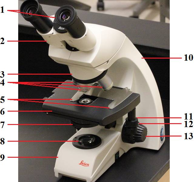
38 diagram of a microscope and label
Label the microscope — Science Learning Hub Label the microscope Add to collection Use this interactive to identify and label the main parts of a microscope. Drag and drop the text labels onto the microscope diagram. eye piece lens coarse focus adjustment high-power objective diaphragm or iris base fine focus adjustment light source stage Download Exercise Tweet Labeling the Parts of the Microscope Labeling the Parts of the Microscope This activity has been designed for use in homes and schools. Each microscope layout (both blank and the version with answers) are available as PDF downloads. You can view a more in-depth review of each part of the microscope here. Download the Label the Parts of the Microscope PDF printable version here.
Microscope, Microscope Parts, Labeled Diagram, and Functions Microscope, Microscope Parts, Labeled Diagram, and Functions What is Microscope? A microscope is a laboratory instrument used to examine objects that are too small to be seen by the naked eye. It is derived from Ancient Greek words and composed of mikrós, "small" and skopeîn,"to look" or "see".
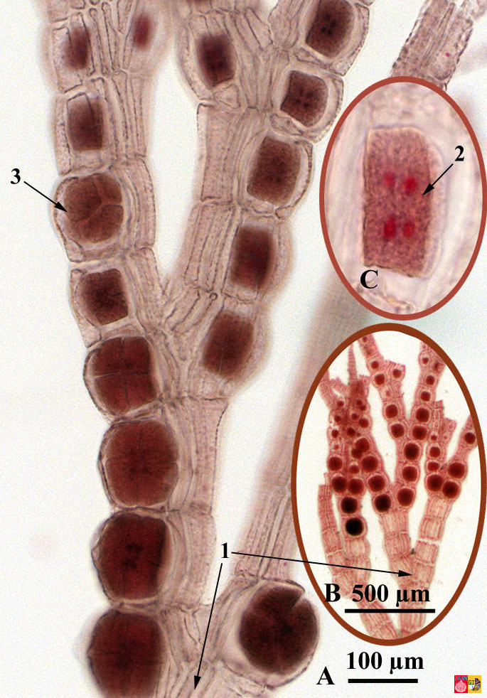
Diagram of a microscope and label
Compound Microscope Parts, Functions, and Labeled Diagram Compound Microscope Parts, Functions, and Labeled Diagram Parts of a Compound Microscope Each part of the compound microscope serves its own unique function, with each being important to the function of the scope as a whole. Fluorescence - Wikipedia Fluorescence is the emission of light by a substance that has absorbed light or other electromagnetic radiation.It is a form of luminescence.In most cases, the emitted light has a longer wavelength, and therefore a lower photon energy, than the absorbed radiation. Microscope Types (with labeled diagrams) and Functions Simple microscope labeled diagram Simple microscope functions It is used in industrial applications like: Watchmakers to assemble watches Cloth industry to count the number of threads or fibers in a cloth Jewelers to examine the finer parts of jewelry Miniature artists to examine and build their work Also used to inspect finer details on products
Diagram of a microscope and label. Simple Microscope - Parts, Functions, Diagram and Labelling Parts of the optical parts are as follows: Mirror - A simple microscope has a plano-convex mirror and its primary function is to focus the surrounding light on the object being examined. Lens - The biconvex lens is placed above the stage and its function is to magnify the size of the object being examined. Microscope Labeling Diagram | Quizlet Start studying Microscope Labeling. Learn vocabulary, terms, and more with flashcards, games, and other study tools. Connective Tissue and Quiz 1 - histology Note at high power that fibrils or fibers of any type cannot be readily observed. Now with slide 28 (make sure your slide is stained with silver; it should say “Ag” on the label!), note how a network of fine black fibrils is present in this same tissue following silver staining. These are reticular fibers, found in skin, muscle and blood ... Neuron under Microscope with Labeled Diagram Neuron under Microscope with Labeled Diagram 31/03/2022 31/03/2022 by anatomylearner The structural and functional unit of the nervous system is the neuron that may easily observe under a light microscope. Neurons may vary considerably in size, shape, and other features.
Stain protocols 2010 - Austin Community College District with the clean microscope slide and using the capillary action of the dye/ slide to spread the India ink across the smear. Refer to the Negative Stain portion of this handout for a diagram. 4. Allow the film to air dry. DO NOT heat or blot dry!!!! Heat will melt the capsule! 5. Saturate the slide with crystal violet for 1 minute. 6. Welcome to Virtual Urchin - University of Washington Major update Apr 2021: All of the activities on the site are now mobile compatible !! Computers are still recommended, and tablets are preferable to phones: please read the Notes at the bottom of this page for details on the latest updates, mobile compatibility and general information about using this site. Microscope Parts and Functions With Labeled Diagram and Functions How ... First, the purpose of a microscope is to magnify a small object or to magnify the fine details of a larger object in order to examine minute specimens that cannot be seen by the naked eye. Here are the important compound microscope parts... Eyepiece: The lens the viewer looks through to see the specimen. Compound Microscope- Definition, Labeled Diagram, Principle, Parts, Uses The optical microscope often referred to as the light microscope, is a type of microscope that uses visible light and a system of lenses to magnify images of small subjects. There are two basic types of optical microscopes: Simple microscopes. Compound microscopes. The term "compound" in compound microscopes refers to the microscope having ...
PDF Label parts of the Microscope: Answers Label parts of the Microscope: Answers Coarse Focus Fine Focus Eyepiece Arm Rack Stop Stage Clip . Created Date: 20150715115425Z ... The Parts of a Microscope (Labeled) Printable - TeacherVision The Parts of a Microscope (Labeled) Printable. This diagram labels and explains the function of each part of a microscope. Use this printable as a handout or transparency to help prepare students for working with laboratory equipment. Grade: Label Microscope Diagram - EnchantedLearning.com Using the terms listed below, label the microscope diagram. arm - this attaches the eyepiece and body tube to the base. base - this supports the microscope. body tube - the tube that supports the eyepiece. coarse focus adjustment - a knob that makes large adjustments to the focus. diaphragm - an adjustable opening under the stage, allowing ... A Study of the Microscope and its Functions With a Labeled Diagram These labeled microscope diagrams and the functions of its various parts, attempt to simplify the microscope for you. However, as the saying goes, 'practice makes perfect', here is a blank compound microscope diagram and blank electron microscope diagram to label. Download the diagrams and practice labeling the different parts of these ...
Life cycle of Polysiphonia (Rhodophyta Red algae)
What is a Bunsen Burner? | Bunsen Burner Parts, Diagram ... Jul 29, 2021 · The Parts of a Bunsen Burner. The entire burner is typically made of metal and it burner sits on the bench via a sturdy base. The main gas inlet at the base of the burner is attached to gas nozzle ...
leica_dm500.jpg
Label Microscope Diagram - EnchantedLearning.com Label Microscope Diagram Using the terms listed below, label the microscope diagram. Inventions and Inventors: arm - this attaches the eyepiece and body tube to the base. base - this supports the microscope. body tube - the tube that supports the eyepiece. coarse focus adjustment - a knob that makes large adjustments to the focus.

PhillipsBiology: Microscope Lab - Vascular plants
Compound Microscope Parts - Labeled Diagram and their Functions - Rs ... There are three major structural parts of a compound microscope. The head includes the upper part of the microscope, which houses the most critical optical components, and the eyepiece tube of the microscope. The base acts as the foundation of microscopes and houses the illuminator. The arm connects between the base and the head parts.
Mitosis Videos
Microscope Labeling - The Biology Corner 1) Start with scanning (the shortest objective) and only use the COARSE knob . Once it is focused… 2) Switch to low power (medium) and only use the COARSE knob . You may need to recenter your slide. Once it is focused.. 3) Switch to high power (long objective).

Parts of a Compound Light Microscope on Vimeo
Microscope Parts, Function, & Labeled Diagram - slidingmotion Microscope parts labeled diagram gives us all the information about its parts and their position in the microscope. Microscope Parts Labeled Diagram The principle of the Microscope gives you an exact reason to use it. It works on the 3 principles. Magnification Resolving Power Numerical Aperture. Parts of Microscope Head Base Arm Eyepiece Lens

Cells Under A Microscope by Jaimarie Nelson
Microscope Labeling - The Biology Corner Students label the parts of the microscope in this photo of a basic laboratory light microscope. Can be used for practice or as a quiz. ... Microscope Labeling . Microscope Use: 15. When focusing a specimen, you should always start with the _____ objective. 16. When using the high power objective, only the _____ knob should be used. 17. The ...

Quia - AP Chapter 18 -Sections 1 and 2-Viral Genetics (basic)
Parts of a microscope with functions and labeled diagram Q. List down the 18 parts of a Microscope. 1. Ocular Lens (Eye Piece) 2. Diopter Adjustment 3. Head 4. Nose Piece 5. Objective Lens 6. Arm (Carrying Handle) 7. Mechanical Stage 8. Stage Clip 9. Aperture 10. Diaphragm 11. Condenser 12. Coarse Adjustment 13. Fine Adjustment 14. Illuminator (Light Source) 15. Stage Controls 16. Base 17.
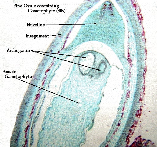
Lab 24 Gymnosperms
PDF Parts of a Microscope Printables - Homeschool Creations Label the parts of the microscope. You can use the word bank below to fill in the blanks or cut and paste the words at the bottom. Microscope Created by Jolanthe @ HomeschoolCreations.net. Parts of a eyepiece arm stageclips nosepiece focusing knobs illuminator stage objective lenses

UNBiology1
Microscope Labeling Practice Diagram | Quizlet Where the microscope slide is placed for viewing. Coarse Focus Knob (Coarse Adjustment) Elevates or lowers the stage a large distance with each turn of the knob. Fine Focus Knob (Fine Adjustment) Elevates or lowers the stage a small distance with each turn or the knob. Base Provides a firm and steady support. Carry with one hand here. Body Tube
Post a Comment for "38 diagram of a microscope and label"