41 label the internal features of stomach and duodenum using the hints if provided.
A&P 2 Lab Practical Final Flashcards - Quizlet Put the following structures of the lower respiratory tract in order from proximal to distal. Label these structures of the upper respiratory system. Label the anterior view of the larynx based on the hints if provided. Place the following words in order to show the pathway oxygen will diffuse across the respiratory membrane Module 3 Study Guide ch 24 ch 25Catrina Greene BIO-169 ... - Course Hero Label the parts of the liver and gallbladder using the hints provided. Label the abdominal contents using the hints provided. Label the sagittal section of the mesenteries. Label the mucous membrane tissue from the stomach using the hints if provided. Correctly organize the events of the defecation reflex.
Anatomical Directional Terms and Body Planes - ThoughtCo These prefixes and suffixes give us hints about the locations of body structures. For example, the prefix (para-) means near or within. The parathyroid glands are located on the posterior side of the thyroid. The prefix epi- means upper or outermost. The epidermis is the outermost skin layer. The prefix (ad-) means near, next to, or toward.

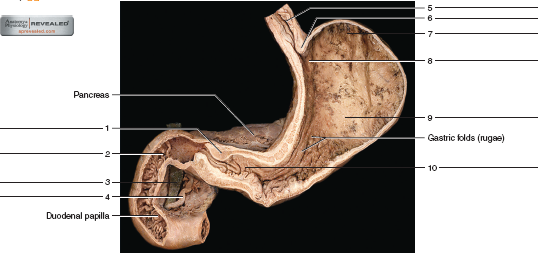
Label the internal features of stomach and duodenum using the hints if provided.
23.3 The Mouth, Pharynx, and Esophagus - Anatomy & Physiology In this section, you will examine the anatomy and functions of the three main organs of the upper alimentary canal—the mouth, pharynx, and esophagus—as well as three associated accessory organs—the tongue, salivary glands, and teeth. The Mouth The cheeks, tongue, and palate frame the mouth, which is also called the oral cavity (or buccal cavity). 007460.pdf - LESSON 5 THE HUMAN BODY In the fifth lesson ... - Course Hero In your stomach, mechanical digestion continues as does chemical digestion, which occurs thanks to hydrochloric acid, pepsin, and mucus. Pepsin breaks down proteins in acidic conditions and mucus lines the stomach to protect it from the acid. Once these juices and the food mix together, the mixture leaving the stomach is referred to as chyme. Digestive lab Flashcards | Quizlet Label the mucous membrane tissue from the stomach using the hints if provided. Label the digestive abdominal contents using the hints if provided. Place the appropriate words and descriptions with the picture with the correct highlighted digestive accessory organ. Label the structures of the posterior thoracic wall using the hints if provided.
Label the internal features of stomach and duodenum using the hints if provided.. The Dogfish Shark—Structure and FUNction! - Carolina.com The stomach's longitudinal folds, called rugae, allow the stomach to expand. Discuss these digestive structures in light of the fact that the shark does not chew its food but instead bites off and swallows large chunks of it. At a J-shaped turn along the digestive tube, the stomach leads into the duodenum. 9789386663542 Maple G05 Evs I (Science) Workbook Term 1 The Maple EVS - I (Science) textbooks and workbooks offer the following features: Interactive content that engages students through a range of open- ended questions that build curiosity and initiate exploration Opportunities for experimentation, analysis and synthesis of ideas and concepts Exposure to locally relevant environmental problem ... Solved Label the internal features of stomach and duodenum - Chegg Label the internal features of stomach and duodenum using the hints if provided. Show transcribed image text Expert Answer 100% (10 ratings) -The esophagus is a muscular tube connecting the throat (pharynx) with the stomach. -The stomach is a muscular organ located on the left side of the upper abdomen. The stomach receives food from the esophagus. 23.1 Overview of the Digestive System - Anatomy & Physiology Muscularis mucosa —This thin layer of smooth muscle is in a constant state of tension, pulling the mucosa of the stomach and small intestine into undulating folds. These folds dramatically increase the surface area available for digestion and absorption. As its name implies, the submucosa lies immediately beneath the mucosa.
Solved Label the internal features of stomach and duodenum | Chegg.com Expert Answer 100% (5 ratings) Explanation - The image shows internal features of stomach and duodenum. * Lower esophageal sphincter Lower esophageal sphincter is bundle of smooth muscles seen at the end of esophagus. * … View the full answer Transcribed image text: Label the internal features of stomach and duodenum using the hints if provided. PDF Cadaver Anatomy Textbook | event.zain latest edition, captured using the most up-to date imaging technologies to ensure excellent visualization of the anatomy. Human Sectional Anatomy Atlas of Body Sections, CT and MRI ... - UnitedVRG Label the abdominal contents using the hints if provided. cadaver. Label the internal features of stomach and duodenum using the hints if provided. Lab 9 Digestive and resp Flashcards - Quizlet stomach Label the various abdominal structures using the hints provided. diaphragm liver pancreas transverse mesocolon duodenum transverse colon jejunum and ileum Label the various abdominal structures using the hints provided. anal canal rectum partial peritoneum greater omentum stomach visceral peritoneum 4.1 Types of Tissues - Anatomy & Physiology Epithelial tissue refers to groups of cells that cover the exterior surfaces of the body, line internal cavities and passageways, and form certain glands. Connective tissue, as its name implies, binds the cells and organs of the body together. Muscle tissue contracts forcefully when excited, providing movement.
Solved Label the histologic features of the jejunum using - Chegg 100% (8 ratings) In the above picture I have labelled the parts of the jejunum. --->The jejunum is the part of small intestine.The j …. View the full answer. Transcribed image text: Label the histologic features of the jejunum using the hints if provided. Epithelium Lacteal Brush border Intestinal gland Paneth cell Villus Goblet cell. Digestive lab Flashcards | Quizlet Label the mucous membrane tissue from the stomach using the hints if provided. Label the digestive abdominal contents using the hints if provided. Place the appropriate words and descriptions with the picture with the correct highlighted digestive accessory organ. Label the structures of the posterior thoracic wall using the hints if provided. Digestive system - Histology The digestive tract (a.k.a alimentary tract ), starts in the oral cavity and continues through the pharynx, to the esophagus, stomach, duodenum, small intestine, large intestine, rectum, and terminates in the anal canal. Food moves along the digestive tract by peristalsis, the rhythmic contractions of the smooth muscle within the walls of the tube. Types of Tissues | Anatomy and Physiology I | | Course Hero Tissue Membranes A tissue membrane is a thin layer or sheet of cells that covers the outside of the body (for example, skin), the organs (for example, pericardium), internal passageways that lead to the exterior of the body (for example, abdominal mesenteries), and the lining of the moveable joint cavities.There are two basic types of tissue membranes: connective tissue and epithelial ...
How to Use the Paleo Diet to Treat Gastroparesis, the “Tired Gut ...
Upper GI | Esophagram | Barium Swallow - Radiologyinfo.org Upper gastrointestinal tract radiography, also called an upper GI, is an x-ray examination of the esophagus, stomach and first part of the small intestine (also known as the duodenum). Images are produced using a special form of x-ray called fluoroscopy and an orally ingested contrast material such as barium.
34 Label The Features Of The Stomach And Nearby Regions - Label Design ...
GI Post Lab Flashcards | Quizlet Label the internal features of stomach and duodenum using the hints if provided. Label the histologic features of the duodenum using the hints if provided. Label the light micrograph of the colon using the hints provided. Label the light micrograph of the liver using the hints provided.
Post a Comment for "41 label the internal features of stomach and duodenum using the hints if provided."