39 label the photomicrograph of the sebaceous gland.
LookWAYup definition of - senses, usage, synonyms, thesaurus. Online Dictionaries: Definition of Options|Tips Solved Label the photomicrograph of the skin and its | Chegg.com Label the photomicrograph of the skin and its accessory structures Epidermis Duct of sebaceous gland Hair follicle Sebaceous gland Show transcribed image text Expert Answer 2. The picture here demonstrates the pseudostratified columnar epithelium.
Label The Photomicrograph Of The Sebaceous Gland : Histochemical ... Label the photomicrograph of the sebaceous gland. These structures embryologically originate from . If the gland become blocked, the sebum can be forced out into the dermis, where it elicits an inflammatory response. Label the photomicrograph of the skin and its accessory structures. Epidermis hair follicle duct of sebaceous gland sebaceous gland.

Label the photomicrograph of the sebaceous gland.
Anime Base Angry : Lerpish On Twitter Working On A Nagatorosan Model In ... Hair sebaceous gland dermis hair follicle epidermis duct of sebaceous. Label the photomicrograph... Crayola Crayon Wrapper Template : Free Printable Crayola Crayon Templates Crayon Template Templates Printable Free Crayola Crayons. Crayola® crayon labels cannot be personalized. Add your name to your crayons or make them kawaii cute with these ... HelenakruwAllison Gland Hari Inspen Is Jadual Kelapa Kuala Kulit Label Lagu Lease Lengkap Malaysia Market Nak Nostalgia of Ondeh Pandan Poland Price Raya Renal Rock Sejarah Selamat Selangor Soalan Staging the Timun Untuk Value wallpaper Which Yang CH 5 Integument - CHAPTER 5 INTEGUMENT Skin(Integument ... - Course Hero Figure 5.2a The main structural features of the skin epidermis. Dermis Stratum spinosum Several layers of keratinocytes unified by desmosomes. Cells contain thick bundles of intermediate filaments made of pre-keratin. Stratum basale Deepest epidermal layer; one row of actively mitotic stem cells; some newly formed cells become part of the more superficial layers.
Label the photomicrograph of the sebaceous gland.. Mandala Pferde Zum Ausmalen / Mandala Ausmalbilder Entspannung ... Hair sebaceous gland dermis hair follicle epidermis duct of sebaceous. Label the photomicrograph... Crayola Crayon Wrapper Template : Free Printable Crayola Crayon Templates Crayon Template Templates Printable Free Crayola Crayons. Crayola® crayon labels cannot be personalized. Add your name to your crayons or make them kawaii cute with these ... Final Exam A&P 1 Flashcards | Quizlet Label the photomicrograph of thin skin Hair shaft, epidermis, dermal root sheath, sebaceous gland, dermis, hair matrix label the structures of the hair follicle Identify the layers of the epidermis with relation to their location and role in keratinization ... the receptors responsible for olfaction are located in the olfactory epithelium Solved Label the photomicrograph of the sebaceous gland. | Chegg.com Experts are tested by Chegg as specialists in their subject area. We review their content and use your feedback to keep the quality high. Transcribed image text: Label the photomicrograph of the sebaceous gland. Sebaceous Gland - an overview | ScienceDirect Topics Sebaceous glands pour their lipid-rich secretions into hair follicles. The glands vary in size: large glands are lobulated. The alveoli of the glands are filled with cells. Near the periphery of the alveoli these are small, generally cuboidal and contain many lipid droplets in their cytoplasm. Occasional mitoses can be found in this layer of cells.
APR 3 Flashcards | Quizlet Merocrine gland 1. Secretes sweat 2. Ducts open directly on surface of skin 3. Functions in temperature regulation Apocrine gland 1. Secretes sweat 2. Ducts open into hair follicles 3. Secretion is influenced by hormones Sebaceous gland 1. Secretes sebum 2. Ducts open into hair follicles 3. Secretion is influenced by hormones Integumentary System HW_answers.docx - Course Hero Hair follicle , sebaceous gland , sweat gland. 11. cells indicated. 12. In the micrograph of a longitudinal section through a nail, identify the parts of a nail and surrounding structures. 13. Label the structures of the fingernail in a lateral view. 14. ... Label the photomicrograph of the sebaceous gland. 20. (Get Answer) - Label The Photomicrograph Of The Skin And ... - Transtutors The Integumentary System A. Anatomy of the skin. Identify and label the photograph of the skin model: epidermis, dermis, dermal papillae, hypodermis, hair follicle, hair bulb, sebaceous gland, sudoriferous gland, duct of sudoriferous gland, pore of... Activity 4 Differentiating Sebaceous and Sweat Glands Microscopically Using the slide thin ... Label The Photomicrograph Based On The Hints Provided / Endocrine Lab ... Distinguish the different types of pituitary cells using the light microscope and electron microscope. Label the photomicrograph based on the hints provided zona fasciculata suprarenal gland zona reticularis capillary medulla dr thomas . Capsule zona glomerulosa zona fasciculata capillaries suprarenal gland fascicle of cells .
Anatomy and Physiology Homework Chapter 6 Flashcards - Quizlet Consider sebaceous and ceruminous glands. Then click and drag each label into the appropriate category based on whether it pertains to sebaceous glands, ceruminous glands, or both.-Usually opens up into a hair follicle-Secretes sebum-Coats guard hairs to improve their performance-Coats the scalp hair with oil-Blockage and infection can cause pimples unit 4 lab.docx - LAB Unit 4 EXERCISE 7: The ... - Course Hero FIGURE 7.2: Photomicrograph of the skin. epidermis (EPI-derm-is) • dermal papillae (puh-PILL-ee) • hypodermis (HY-poh-der-mis) • papillary (PAP-il-lary) layer of dermis • reticular layer of dermis 1. Dermal Papillae 2. Epidermis 3. Papillary layer of dermis 4. Reticular layer of dermis 5. Hypodermis Sebaceous Adenoma: Background, Pathophysiology, Etiology Background. Sebaceous glands are holocrine, oil-producing glands present in the dermis of mammalian skin. Sebaceous glands are usually attached to hair follicles and are part of a complex skin-adnexal unit known as the folliculoapocrine (sweat) apparatus. The glands are present over the entire body, with the exception of the palms of the hands ... Solved > Question 31 points Label the photomicrograph of ... - ScholarOn 31 points Label the photomicrograph of thin skin. Hair Follicle Hair Dermis Sebaceous gland Duct of sebaceous gland Reset zoom
35 Label The Photomicrograph Of Thick Skin - Label Design Ideas 2020
Junqueira's Basic Histology Text and Atlas, 14th Edition Enter the email address you signed up with and we'll email you a reset link.

Integumentary System - Gland Development - Embryology
Fohlen Pferde Ausmalbilder Zum Ausdrucken / Pferde Malvorlagen Fohlen ... Hair sebaceous gland dermis hair follicle epidermis duct of sebaceous. Label the photomicrograph... Crayola Crayon Wrapper Template : Free Printable Crayola Crayon Templates Crayon Template Templates Printable Free Crayola Crayons. Crayola® crayon labels cannot be personalized. Add your name to your crayons or make them kawaii cute with these ...

HISTOLOGY, Integument, Pigmented (thin) skin
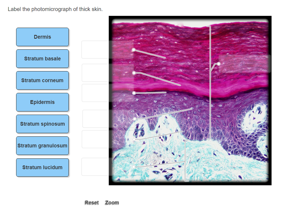
Chapter 6 - Labeling Parts of Skin - sweat gland blood... Labeling Parts of the Skin Identify the layers of skin. dermis stratum basale stratum spinosum stratum lucidum stratum corneum stratum granulosum basement membrane Identify the parts of this photomicrograph of skin: sebaceous gland hair shaft hair follicle dermis epidermis End of preview. Want to read all 2 pages? Upload your study docs or become a

PPT - Assessment of Clients with Integumentary Disorders PowerPoint ...
Figure 7.4 Photomicrograph of the skin and accessory ... - Quizlet Start studying Figure 7.4 Photomicrograph of the skin and accessory structures. Learn vocabulary, terms, and more with flashcards, games, and other study tools.

31 Label The Photomicrograph Of The Sebaceous Gland. - Label Ideas 2021
Bio Lab Chapter 6 Quiz Flashcards | Quizlet -hair follicle and sebaceous gland Identify the type of tissue that composes the epidermis of the skin. stratified squamous epithelial tissue Identify the structures of the dermis. dense connective tissue with fibers oriented in many directions dense irregular loose connective tissue characterized by long, thin dark fiber areolar tissue
Biology Archive | January 15, 2017 | Chegg.com
Anatomy, Skin (Integument), Epidermis - NCBI Bookshelf The exocrine functions of the skin are by way of the sweat and sebaceous glands. Another important role of the skin is a sensation to touch, heat, cold, and pain by the actions of the nociceptors. The general appearance, turgor, and other qualities also give insight into the general health of the body.

Human Scalp Shows Hair Follicle Hair Shaft Sebaceous Glands Dermis ...
Answered: • hair bulbs • hair follicle • hair… | bartleby Solution for • hair bulbs • hair follicle • hair root • papilla of hair • sebaceous gland 1 1 2 2 3 3 4 5 4 5 10x Courtesy Michael Ross, University of Florida
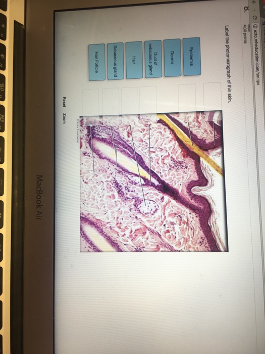
Solved: Label The Photomicrograph Of Thin Skin. | Chegg.com
PDF Name the Condition - Dr. Scott Croes' Website Identify the following structures:nail fold (NF), the matrix region of the nail root (M), the nail bed (NB), nail proper (N), as well as eponychium (Ep) and hyponychium (Hy). A portion of the distal phalanx is also visible showing the secondary ossification centre (2o) and epiphysis.

29 Label The Photomicrograph Of The Sebaceous Gland. - Modern Labels ...
A&P 1 Exercise_7 Activity 1 & 2 & RYK and UYK.docx - Course Hero Apocrine sweat Gland Label the photomicrograph in Figure 7.4. 1. Sebaceous glands 2. Hair follicle 3. Hair root 4. Hair bulb 5. Papilla of hair ... Sebaceous glands 5. Secretes sebum onto hair and skin. Nail body 6. Part of nail that is visible. Free edge 7. Part of nail that extends beyond digit. Hair bulb 8.
Post a Comment for "39 label the photomicrograph of the sebaceous gland."