45 draw and label the microscope
Parts of the Microscope with Labeling (also Free Printouts) Parts of the Microscope with Labeling (also Free Printouts) A microscope is one of the invaluable tools in the laboratory setting. It is used to observe things that cannot be seen by the naked eye. Table of Contents 1. Eyepiece 2. Body tube/Head 3. Turret/Nose piece 4. Objective lenses 5. Knobs (fine and coarse) 6. Stage and stage clips 7. Aperture Microscope Labeling - The Biology Corner Microscope Labeling Microscope Labeling 15. When focusing a specimen, you should always start with the _____________ objective. 16. When using the high power objective, only the _______________ knob should be used. 17. The type of microscope used in most science classes is the ______________ microscope. 18.
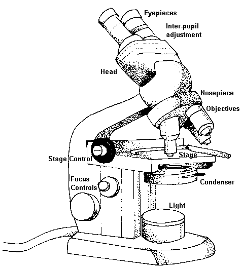
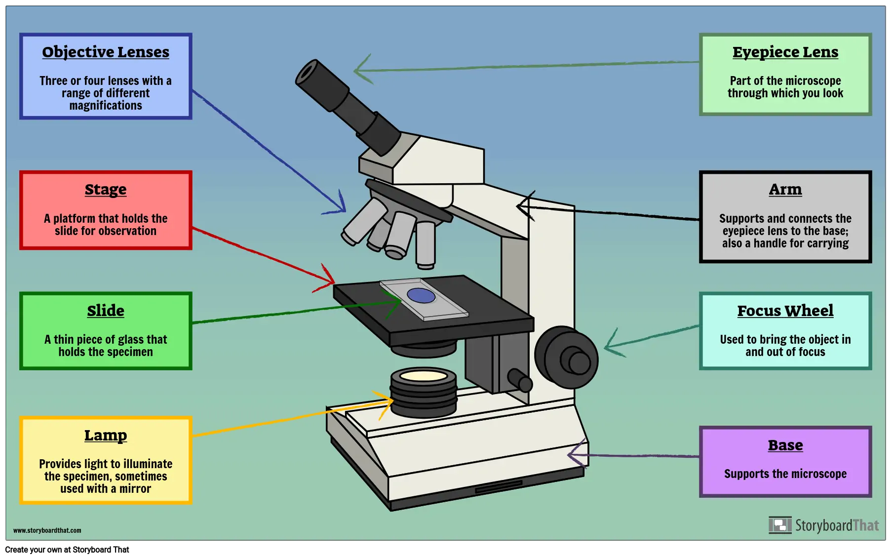
Compound Microscope Parts, Functions, and Labeled Diagram Common compound microscope parts include: Eyepiece (ocular lens) with or without Pointer: The part that is looked through at the top of the compound microscope. Eyepieces typically have a magnification between 5x & 30x. Monocular or Binocular Head: Structural support that holds & connects the eyepieces to the objective lenses.

Draw and label the microscope
Microscope, Microscope Parts, Labeled Diagram, and Functions The proper way to focus a microscope is to start with the lowest power objective lens and crank the lens down as close to the specimen as possible without touching it while looking from the eye piece. Examine the image through the eyepiece lens and focus only upward until it is sharp. If you can't get it to focus, go through the process again. Compound Microscope Parts - Labeled Diagram and their Functions - Rs ... Two adjustment knobs are used to focus the microscope: fine focus knob and coarse focus knob. Both knobs can move the stage up and down. You should use the coarse focus knob to bring the specimen into approximate or near focus. Then you use the fine focus knob to sharpen the focus quality of the image. Label the Microscope Diagram | Download Scientific Diagram Label the Microscope Diagram ... Also, the class provides students with an opportunity to perform useful assays, draw conclusions from their results, and discuss possible extensions of the study ...
Draw and label the microscope. Compound Microscope- Definition, Labeled Diagram, Principle, Parts, Uses The naked eye can now view the specimen at magnification 400 times greater and so microscopic details are revealed. Alternatively, the magnification of the compound microscope is given by: m = D/ fo * L/fe where, D = Least distance of distinct vision (25 cm) L = Length of the microscope tube fo = Focal length of the objective lens PDF Label parts of the Microscope: Answers Label parts of the Microscope: Answers Coarse Focus Fine Focus Eyepiece Arm Rack Stop Stage Clip . Created Date: 20150715115425Z ... Draw the label parts of microscope? - Answers Draw the label parts of microscope? Add Fluorescence to EPhys, Pclamp Ca2+, Fluo-dyes, FRET, Na2+, pH. What is a Compound Microscope? - BYJUS A compound microscope is defined as. A microscope with a high resolution and uses two sets of lenses providing a 2-dimensional image of the sample. The term compound refers to the usage of more than one lens in the microscope. Also, the compound microscope is one of the types of optical microscopes. The other type of optical microscope is a ...
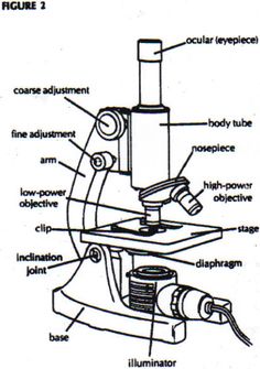
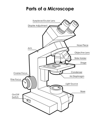
Labeling the Parts of the Microscope | Microscope activity, Science ... Description Worksheet identifying the parts of the compound light microscope. Answer key: 1. Body tube 2. Revolving nosepiece 3. Low power objective 4. Medium power objective 5. High power objective 6. Stage clips 7. Diaphragm 8. Light source 9. Eyepiece 10. Arm 11. Stage 12. Coarse adjustment knob 13. Fine adjustment knob 14. Base S Light Microscope- Definition, Principle, Types, Parts, Labeled Diagram ... A light microscope is a biology laboratory instrument or tool, that uses visible light to detect and magnify very small objects and enlarge them. They use lenses to focus light on the specimen, magnifying it thus producing an image. The specimen is normally placed close to the microscopic lens. Microscope Activity - MICROBIOLOGY - 1... Draw a compound microscope. 2 ... A microscope is said to be parfocal when an ocular lens of a microscope don't lose focus when the objective lenses in use are adjusted while trying to find a better view for the sample. It is useful when the microscope have parfocal feature so the user or the phlebotomist don't need to adjust the focus when changing the power of ... Microscope Parts and Functions A standard microscope has three, four, or five objective lenses that range in power from 4X to 100X. When focusing the microscope, be careful that the objective lens doesn't touch the slide, as it could break the slide and destroy the specimen. Specimen or slide: The specimen is the object being examined.
Compound Microscope - Diagram (Parts labelled), Principle and Uses What are the 13 parts of a microscope? 1. Eyepiece 2. Eyepiece Tube 3. Objective Lens 4. Stage 5. Stage Clips 6. Nosepiece 7. Fine and Coarse Focus knobs 8. Illuminator 9. Aperture 10. Iris Diaphragm 11. Condenser 12. Condenser Focus Knob 13. The Rack stop Q 5. What are the 11 parts of a compound microscope? Microscope Drawing Easy with Label - YouTube In this video I go over a microscope drawing that is easy with label. There is a blank copy at the end of the video to review on your own. A great way to s... Microscope Drawing: How to Sketch Microscope Slides How to Draw Microscope Slides Organize and orient your field of view: To begin, draw a circle as large as possible with a pencil. An 8.5 x 11-inch piece of paper is good size for beginners. The circle represents what you see through the eyepiece of the microscope. Using thin lines, divide the circle into quarters in order to organize the picture. Label the microscope — Science Learning Hub All microscopes share features in common. In this interactive, you can label the different parts of a microscope. Use this with the Microscope parts activity to help students identify and label the main parts of a microscope and then describe their functions. Drag and drop the text labels onto the microscope diagram.
microscopy how a microscope works magnification calculations ...
Simple Microscope - Parts, Functions, Diagram and Labelling The metal stand is an important part for it serves as the support of the entire microscope's part and provides stability to other parts. Stage - The stage of the microscope is a metal plate that is rectangular in shape and fitted to the vertical rod. It comes with a hole in the center that enables the light to pass from below.

Living Environment Course
How to Sketch a Microscope Slide - Identifying and Sketching Cell ... How to Sketch a Microscope Slide Identifying Cell Structures and Adding Dynamic Elements. Learning how to sketch a microscope slide requires an open-mind, patience and a willingness to learn the basic drawing principles of perspective, size, shape and negative space.. Sketching specimens will provide you with a better understanding, as you study the intricacies of the image you see through the ...

how to draw microscope step by step slow and medium speed
A Study of the Microscope and its Functions With a Labeled Diagram The camera present within the microscope captures images to reveal the finer details of the specimen. This microscope can zoom and view the density of a specimen until it is only a micrometer thick and has a magnification ranging between 1,000 - 250,000x on the fluorescent screen. This microscope needs a computer software to yield precise results.
Cells and Microscopes LO:- use and label a microscope - draw ...
Microscope Drawing Teaching Resources | Teachers Pay Teachers This GEEKS lab sheet gives students room to draw three specimens. It includes labels for the Specimen Name, Magnification, Observations/ Challenges. The last label gives students an opportunity to write down something interest them may observe, or record challenges they had while trying to observe their specimens.

Schematic drawing of the atomic force microscope. | Download ...
Labeling the Parts of the Microscope Labeling the Parts of the Microscope This activity has been designed for use in homes and schools. Each microscope layout (both blank and the version with answers) are available as PDF downloads. You can view a more in-depth review of each part of the microscope here. Download the Label the Parts of the Microscope PDF printable version here.

Compound Microscope Parts – Labeled Diagram and their ...
Answered: 2. Draw and label a microscopic… | bartleby Solution for 2. Draw and label a microscopic presentation of: Positive Stain (indicate stain used) Negative stain (Indicate the stain used)

Parts of a Microscope - TessShaheenmicroscopy
Microscope Diagram Labeled, Unlabeled and Blank | Parts of a Microscope ... Both versions contain high resolution images to clearly illustrate the different parts of the microscope as well as other graphics. Along with a full microscope diagram to label, each part is examined in more detail to properly illustrate each component, its use and function. Click the PREVIEW for a closer look at the PowerPoint slides…

Old microscope color sketch engraving vector illustration ...
How To Draw A Microscope - YouTube Today, we're learning how to draw a cool microscope!👩🎨 JOIN OUR ART HUB MEMBERSHIP! VISIT 🎨 VISIT OUR AMAZON ART SUPPLY S...
Label Parts Of Microscope - ClipArt Best
Microscope Parts, Function, & Labeled Diagram - slidingmotion A microscope is a laboratory instrument used to examine very small or micro-objects such as cells and microorganisms that are not able seen by the naked eye. What are the parts of the Microscope? Head Arm Base Eyepiece Lens Eyepiece Tube Objective Lenses Microscope Illuminator Stage and Stage Clips Microscope Nosepiece Rack Stop Condenser Lens

Instruction/ Situation: (MDL) Draw and label the parts of the ...
Microscope Labeling Diagram | Quizlet Unit 2 Lesson 5 - Punnett Squares and Pedigrees. 4 terms. PGFry210. Unit 2 Lesson 4 - Heredity. 9 terms. PGFry210. Upgrade to remove ads. Only $2.99/month.

Parts of a Microscope - SmartSchool Systems
Label the Microscope Diagram | Download Scientific Diagram Label the Microscope Diagram ... Also, the class provides students with an opportunity to perform useful assays, draw conclusions from their results, and discuss possible extensions of the study ...

Light Microscope PNG - light-microscope-e compound-light ...
Compound Microscope Parts - Labeled Diagram and their Functions - Rs ... Two adjustment knobs are used to focus the microscope: fine focus knob and coarse focus knob. Both knobs can move the stage up and down. You should use the coarse focus knob to bring the specimen into approximate or near focus. Then you use the fine focus knob to sharpen the focus quality of the image.

Labeling the Parts of the Microscope | Microscope World Resources
Microscope, Microscope Parts, Labeled Diagram, and Functions The proper way to focus a microscope is to start with the lowest power objective lens and crank the lens down as close to the specimen as possible without touching it while looking from the eye piece. Examine the image through the eyepiece lens and focus only upward until it is sharp. If you can't get it to focus, go through the process again.
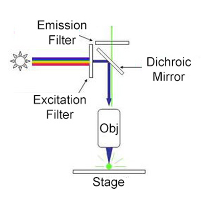
Fluorescence Microscopy - Explanation and Labelled Images ...

Microscope Drawing posted by Samantha Walker
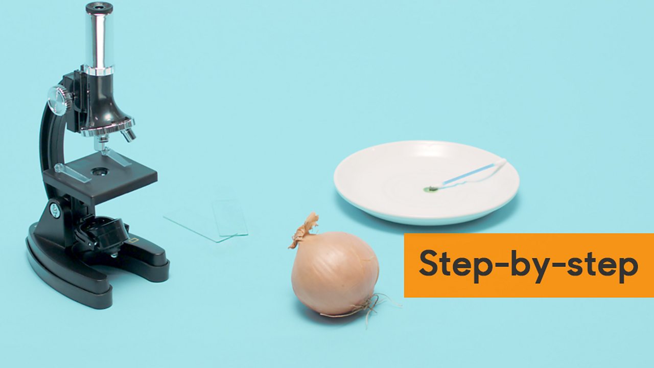
How to observe cells under a microscope - Living organisms ...
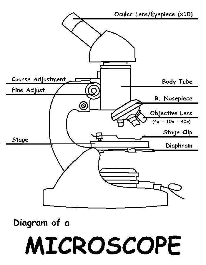
Diagram of a Microscope by ScienceDoodles on DeviantArt
Glossary of terms used in microscopy – Quekett Microscopical Club

Microscope Parts and Functions

China Low price for Lung Model - Lab pathological microscope ...

Label the Microscope Diagram | Download Scientific Diagram

Simple Microscope - Diagram (Parts labelled), Principle ...

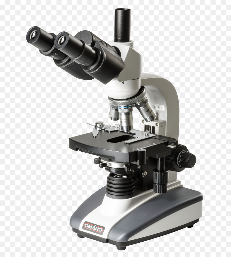
mikroskop olympus seri cx23 di labstor | Tokopedia

How to Draw a Microscope - Really Easy Drawing Tutorial

Label Microscope Diagram - EnchantedLearning.com

2000X Optical Metal Microscope for Adults Kids Students, Two ...

Parts of a microscope with functions and labeled diagram
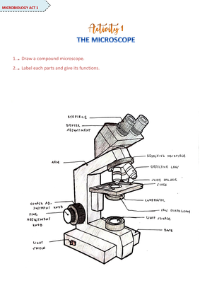
Microscope Activity - MICROBIOLOGY - 1... Draw a compound ...
Microscope, Microscope Parts, Labeled Diagram, and Functions
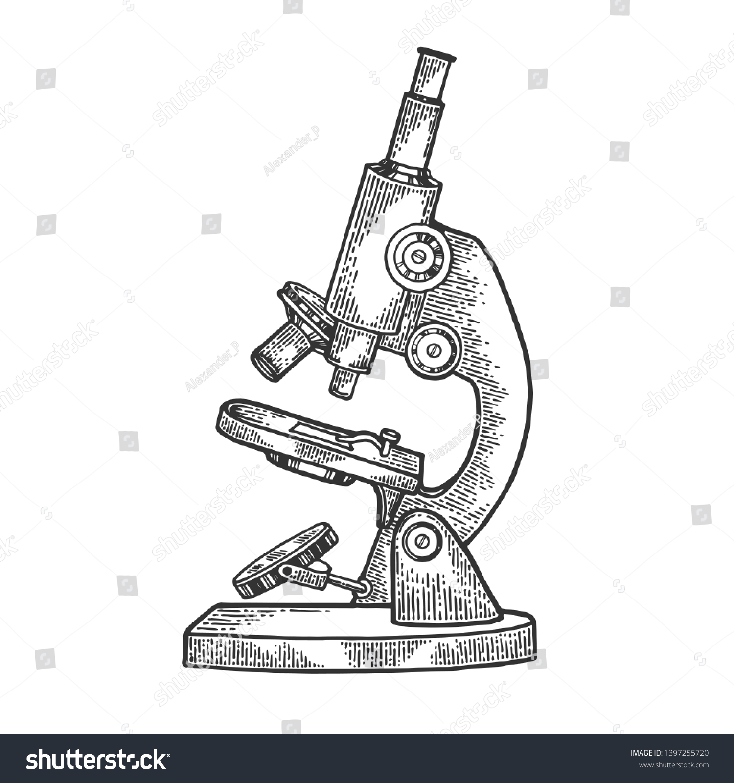
Old Microscope Sketch Engraving Vector Illustration Stock ...

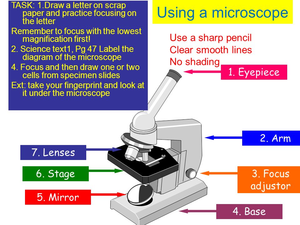
Label the numbered parts of the microscope - ppt download
compound and stereo microscope - Clip Art Library

Collection Of Free Microscopes Drawing Label Clipart ...

Parts Of A Microscope Drawing Grade - Gr 9 Natural Science ...
Cahaya, Mikroskop, Mikroskop Optik gambar png

Draw and label the parts of the microscope. Draw it in your ...
Microscope Diagram Labeled, Unlabeled and Blank | Parts of a ...

Cytology. Cytology. radiation used to illuminate the specimen ...

Microscope Labeling Diagram | Quizlet

List: Parts of a Microscope and their Function | Pathwooded
Parts of a Microscope Labeling Activity
Free Microscope Drawing, Download Free Microscope Drawing png ...

in a long bond paper draw and label the parts of a compound ...

Parts of a Compound Microscope and Their Functions
1.5: Microscopy - Biology LibreTexts
Post a Comment for "45 draw and label the microscope"