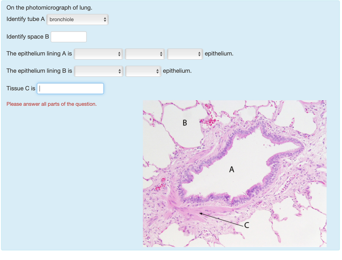
40 label the photomicrogram of the lung
Labeled Diagram of the Human Lungs - Bodytomy Given below is a labeled diagram of the human lungs followed by a brief account of the different parts of the lungs and their functions. Each lung is enclosed inside a sac called pleura, which is a double-membrane structure formed by a smooth membrane called serous membrane. Solved Label the photomicrogram of the lung Alveolar duct - Chegg Expert Answer 100% (1 rating) A.Alveolar duct. B.SMALL BRANCH OF PULMONARY A … View the full answer Transcribed image text: Label the photomicrogram of the lung Alveolar duct Alveolus Small branch of pulmonary a. bronchiole Alveolar sac Reset Zoom < Prev 22 of 40::: Next > 20 Previous question Next question
Lab 2: Microscopy and the Study of Tissues - UW-La Crosse 1. Introduction to histology (Part 1) Tissues are composed of similar types of cells that work in a coordinated fashion to perform a common task, and the study of the tissue level of biological organization is histology. Four basic types of tissues are found in animals. Epithelium is a type of tissue whose main function is to cover and protect ...

Label the photomicrogram of the lung
Lung Anatomy, Function, and Diagrams - Healthline The lungs begin at the bottom of your trachea (windpipe). The trachea is a tube that carries the air in and out of your lungs. Each lung has a tube called a bronchus that connects to the trachea ... Photomicrograph of the Lung Quiz - PurposeGames.com Games by same creator. 2p Image Quiz. 8p Image Quiz. Reproductive Systems 2 games. The Integumentary System. The Nervous System & Senses. The Digestive System. The Human Body-- An Orientation 7 games. The Microscope 2 games. Histology of trachea and lung - SlideShare 1. HISTOLOGY OF TRACHEA AND LUNG Dr.ushakannan,Asst.professor. 2. RESPIRATORY SYSTEM Conducting Part- responsible for passage of air and conditioning of the inspired air. Examples:nasal cavities,pharynx, trachea, bronchi and their intrapulmonary continuations. Respiratory Part-involved with the exchange of oxygen and carbondioxide between blood ...
Label the photomicrogram of the lung. quizlet.com › 159345340 › ap-2-lab-unit-2-flash-cardsA&P 2 Lab Unit 2 Flashcards | Quizlet Label the photomicrogram of the lung. Identify the cartilaginous anatomical structures shown in the posterior view of the superior portion of the lower respiratory system. Place the following structures with the appropriate anatomical region. Labels maybe be placed in more than one category. quizlet.com › 548952862 › unit-6-flash-cardsUnit 6 Flashcards | Quizlet Label the photomicrogram of the trachea. Label the anterior view of the larynx based on the hints if provided. Place the different partial pressure of gases with the appropriate location. A&P 139 Chapter 19 Flashcards | Quizlet The pharynx is an enlargement at the top of the trachea that houses the vocal cords. the contraction of the diaphragm. true false out of the lungs, called expiration Inspiration begins with: As alveolar volume increases, alveolar pressure decreases. Air moves from areas of low pressure to areas of high pressure until an equilibrium is reached. Label Lungs Diagram Printout - Enchanted Learning Read the definitions below, then label the lung anatomy diagram. Extra Information Word Bank bronchial tree: the system of airways within the lungs, which bring air from the trachea to the lung's tiny air sacs (alveoli). cardiac notch: the indentation in the left lung that provides room for the heart. diaphragm: a muscular membrane under the lungs.
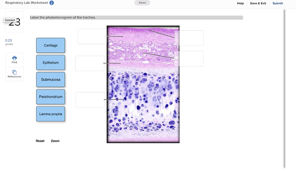
A&P 2 Lab Unit 2 Flashcards | Quizlet Image: Label the photomicrogram of the lung. Identify the cartilaginous anatomical structures shown in the posterior view of the superior ... A&P 2 Lab Unit 2 Flashcards | Quizlet WebStudy with Quizlet and memorize flashcards containing terms like Identify the anatomical structures shown in the anterior view of the superior portion of the lower respiratory system., Put the following layers of the trachea in order from superficial to deep., Label the structures of the upper respiratory system. and more. Label The Photomicrograph Of The Lung : Anatomy Physiology Tissue The ... Label the photomicrogram of the lung segmental branch of pulmonary a. Light micrograph of lung tissue (click to show / hide labels). Collective name for the multiple branches of the bronchi and . (b) a micrograph shows the alveolar structures within lung tissue. Abnormal cells grow and can form tumors. Label the photomicrogram of the trachea. - en.ya.guru The trachea is known to be a kind of long tube that links the human larynx (voice box) to that of their bronchi. Note that the bronchi is one that send air to a person's lungs and the trachea is known to be an essential part of man's respiratory system. Hence, The Label of the photomicrogram of the trachea is given in the image attached.
How to draw and label a lung | step by step tutorial - YouTube A beautiful drawing of Lung. And it will teach you to draw the lung very easily. Watch the video and please be kind enough to thumbs up my videos. And I will... Solved Label the photomicrogram of the lung Segmental branch | Chegg.com Question: Label the photomicrogram of the lung Segmental branch of pulmonary a. Interalveolar wall Alveolar macrophage Reset Zoom < Prev 19 40 , Next > 2 3 4 5 6 This problem has been solved! You'll get a detailed solution from a subject matter expert that helps you learn core concepts. See Answer Show transcribed image text Expert Answer The Lungs | Anatomy and Physiology II - Lumen Learning The right lung is shorter and wider than the left lung, and the left lung occupies a smaller volume than the right. The cardiac notch is an indentation on the surface of the left lung, and it allows space for the heart (Figure 1). The apex of the lung is the superior region, whereas the base is the opposite region near the diaphragm. Anatomy of the Lung | SEER Training - National Cancer Institute Anatomy of the Lung. The lungs are the major organs of the respiratory system, and are divided into sections, or lobes.The right lung has three lobes and is slightly larger than the left lung, which has two lobes.. The lungs are separated by the mediastinum.This area contains the heart, trachea, esophagus, and many lymph nodes. The lungs are covered by a protective membrane known as the pleura ...
Label the lungs | Biology - Quizizz Label the lungs DRAFT. 25 minutes ago by. 15c1rscot_00844. K - Professional development. Biology, Science. Played 0 times. 0 likes. 0% average accuracy. 0. Save. Edit. Edit. Print; Share; Edit; Delete; Report an issue; Live modes. Start a live quiz . Classic . Students progress at their own pace and you see a leaderboard and live results.
Answered: Which structure is highlighted? | bartleby Transcribed Image Text:Label the photomicrogram of the lung. Conducting bronchiole Alveolar duct Alveolar sac Small branch of pulmonary a.
Photomicrography Definition & Meaning - Merriam-Webster The meaning of PHOTOMICROGRAPH is a photograph of a microscope image.
Photomicrogram Definition & Meaning - Merriam-Webster The meaning of PHOTOMICROGRAM is photomicrograph. Love words? You must — there are over 200,000 words in our free online dictionary, but you are looking for one that's only in the Merriam-Webster Unabridged Dictionary.. Start your free trial today and get unlimited access to America's largest dictionary, with:. More than 250,000 words that aren't in our free dictionary
Label The Photomicrograph Of The Lung : 4 Chloro Dl Phenylalanine ... Label the photomicrogram of the lung segmental branch of pulmonary a. Relative amounts of glands, cartilage, smooth muscles and connective tissue fibers present in the wall of the tubes. Photomicrographs of bronchioles and pulmonary alveoli of giant anteater (myrmecophaga tridactyla). Learn what you need to know about lung cancer.
Can you label the lungs? Quiz - PurposeGames.com Labeling the lungs. This online quiz is called Can you label the lungs?. It was created by member birdb08 and has 9 questions. It is currently featured in 8 tournaments.
Labeled diagram of the lungs/respiratory system. - SERC Labeled diagram of the lungs/respiratory system. Image 37789 is a 1125 by 1408 pixel PNG Uploaded: Jan10 14 Last Modified: 2014-01-10 12:15:34 Permanent URL: The file is referred to in 1 page Airborne Microbes
Respiratory APR Flashcards | Quizlet Respiratory APR. Term. 1 / 151. ala of nose. Click the card to flip 👆. Definition. 1 / 151.
label the lungs worksheet - TeachersPayTeachers Science worksheets: Label parts of the human lungs. by. Science Workshop. 5.0. (1) $1.25. $1.24. Word Document File. Science worksheets: Label parts of the human lungs2 VERSIONS OF WORKSHEET (Worksheet with a word bank & Worksheet with no word bank)Students have to parts of the human lungs (Alveoli , Bronchioles, Bronchi, Trachea)Worksheet ...
A&P 2 Lab Practical Final Flashcards | Quizlet (Respiratory) Larynx Label the photomicrogram of the lung. Put the following structures of the lower respiratory tract in order from proximal to distal. Label these structures of the upper respiratory system. Label the anterior view of the larynx based on the hints if provided.
Label the photomicrogram of the trachea. - Brainly.com The trachea is known to be a kind of long tube that links the human larynx (voice box) to that of their bronchi. Note that the bronchi is one that send air to a person's lungs and the trachea is known to be an essential part of man's respiratory system. Hence, The Label of the photomicrogram of the trachea is given in the image attached.
Matching and anatomical labeling of human airway tree - PMC Matching of corresponding branchpoints between two human airway trees, as well as assigning anatomical names to the segments and branchpoints of the human airway tree, are of significant interest for clinical applications and physiological studies. In the past these tasks were often performed manually due to the lack of automated algorithms ...
Histology of trachea and lung - SlideShare 1. HISTOLOGY OF TRACHEA AND LUNG Dr.ushakannan,Asst.professor. 2. RESPIRATORY SYSTEM Conducting Part- responsible for passage of air and conditioning of the inspired air. Examples:nasal cavities,pharynx, trachea, bronchi and their intrapulmonary continuations. Respiratory Part-involved with the exchange of oxygen and carbondioxide between blood ...
Photomicrograph of the Lung Quiz - PurposeGames.com Games by same creator. 2p Image Quiz. 8p Image Quiz. Reproductive Systems 2 games. The Integumentary System. The Nervous System & Senses. The Digestive System. The Human Body-- An Orientation 7 games. The Microscope 2 games.
Lung Anatomy, Function, and Diagrams - Healthline The lungs begin at the bottom of your trachea (windpipe). The trachea is a tube that carries the air in and out of your lungs. Each lung has a tube called a bronchus that connects to the trachea ...


Post a Comment for "40 label the photomicrogram of the lung"