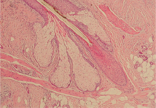
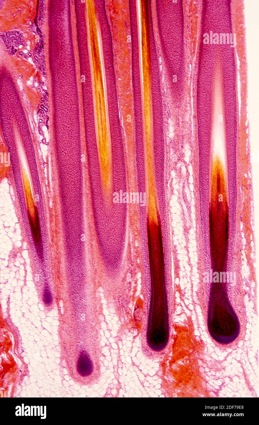
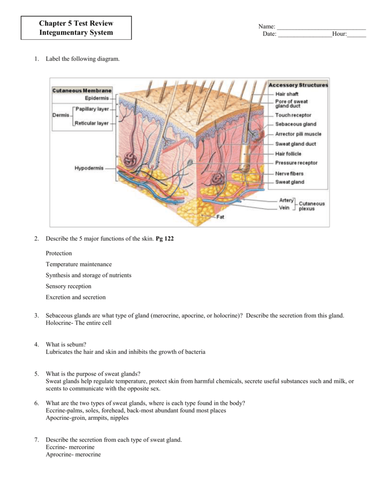
44 label the photomicrograph of the skin and its accessory structures
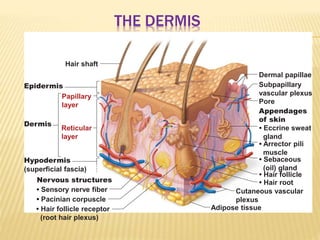
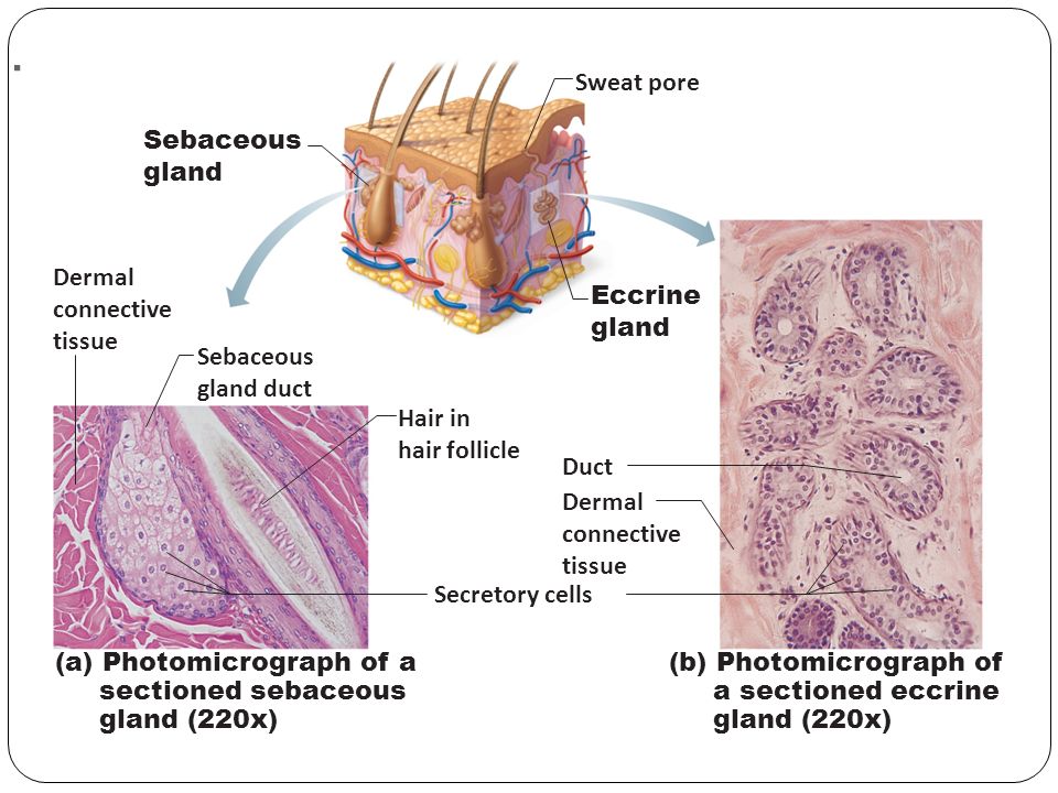
5.1 Layers of the Skin - Anatomy and Physiology 2e - OpenStax The skin and its accessory structures make up the integumentary system, which provides the body with overall protection. The skin is made of multiple layers of cells and tissues, which are held to underlying structures by connective tissue ( Figure 5.2 ). The deeper layer of skin is well vascularized (has numerous blood vessels). Solved Label the photomicrograph of the skin and its - Chegg Label the photomicrograph of the skin and its accessory structures Epidermis Duct of sebaceous gland Hair follicle Sebaceous gland This problem has been solved! You'll get a detailed solution from a subject matter expert that helps you learn core concepts. See Answer
Photomicrograph Definition & Meaning - Merriam-Webster The meaning of PHOTOMICROGRAPH is a photograph of a microscope image. Recent Examples on the Web Delicate hooks on the agrimony seed in this photomicrograph snare the fur of passing animals (or your socks) to move to greener pastures. — Discover Magazine, 29 June 2010 This photomicrograph reveals the disposition of axons that regulate the contraction of certain muscles.
Label the photomicrograph of the skin and its accessory structures
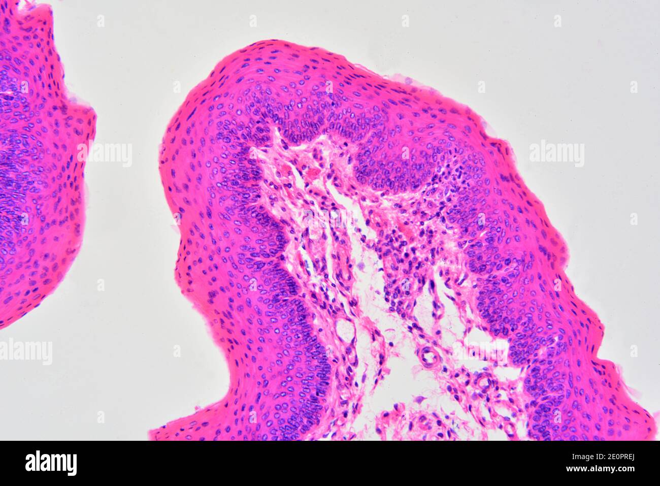
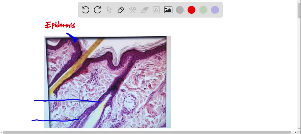
Accessory Structures of the Skin - Anatomy & Physiology Accessory structures of the skin include hair, nails, sweat glands, and sebaceous glands. Hair is made of dead keratinized cells, and gets its color from melanin pigments. Nails, also made of dead keratinized cells, protect the extremities of our fingers and toes from mechanical damage. Layers of the Skin | Anatomy and Physiology I - Lumen Learning The skin is composed of two main layers: the epidermis, made of closely packed epithelial cells, and the dermis, made of dense, irregular connective tissue that houses blood vessels, hair follicles, sweat glands, and other structures. Beneath the dermis lies the hypodermis, which is composed mainly of loose connective and fatty tissues. Structure and Function of Skin | Biology for Majors II - Lumen Learning The skin and its accessory structures make up the integumentary system, which provides the body with overall protection. The skin is made of multiple layers of cells and tissues, which are held to underlying structures by connective tissue (Figure 1). The deeper layer of skin is well vascularized (has numerous blood vessels).
Label the photomicrograph of the skin and its accessory structures. Figure 7.4 Photomicrograph of the skin and accessory structures - Quizlet Science Biology Anatomy Figure 7.4 Photomicrograph of the skin and accessory structures 3.0 (2 reviews) + − Flashcards Learn Test Match Created by dbonner6986 Terms in this set (5) Term Hair Bulb Definition contains the papilla of the hair and the matrix Location Term Papilla of hair Definition AP Lab Midterm - APR 4 Flashcards | Quizlet label the photomicrograph of the skin and its accessory structures epidermis, duct, sebaceous, arrector, sweat, hair second highlighted layer of skin dermis Label the photomicrograph of thin skin. epidermis, hair, duct, sebaceous, hair, dermis Label the structures of the fingernail in a lateral view. (top to bottom) nail body, free edge, lateral 5.2 Accessory Structures of the Skin - OpenStax Accessory structures of the skin include hair, nails, sweat glands, and sebaceous glands. These structures embryologically originate from the epidermis and can extend down through the dermis into the hypodermis. Hair Hair is a keratinous filament growing out of the epidermis. It is primarily made of dead, keratinized cells. Network Rail admits delays will get worse in the next five years More misery for British train passengers as Network Rail bosses admit delays will get WORSE in the next five years due to rising costs and funding. Network Rail will reportedly have to cut their ...
Photomicrography | biology | Britannica photomicrography, photography of objects under a microscope. Such opaque objects as metal and stone may be ground smooth, etched chemically to show their structure, and photographed by reflected light with a metallurgical microscope. Biological materials may be killed, dyed so that their structure can be seen, and mounted on glass slides for photographing by transmitted light using ordinary ... Label The Photomicrograph Of The Skin And Its Accessory Structures ... Key Terms Matching 1. epidermis 2. keratin 3. hair bulb A. portion of hair below skin surface B. secretes oil C. light-colored, half-moon shape at base of nail D. detects fine touch and pressure E. only living layer of the epidermis F. protein giving... Posted 11 months ago Q: Solved Label the Photomicrograph of the skin and its - Chegg Label the Photomicrograph of the skin and its accessory structures. Question: Label the Photomicrograph of the skin and its accessory structures. This problem has been solved! See the answer Show transcribed image text Expert Answer 100% (3 ratings) Transcribed image text: Label the Photomicrograph of the skin and its accessory structures. 5.2 Accessory Structures of the Skin - Anatomy & Physiology Chapter Review. Accessory structures of the skin include hair, nails, sweat glands, and sebaceous glands. Hair is made of dead keratinized cells, and gets its color from melanin pigments. Nails, also made of dead keratinized cells, protect the extremities of our fingers and toes from mechanical damage. Sweat glands and sebaceous glands produce ...
Micrograph - Wikipedia A micrograph or photomicrograph is a photograph or digital image taken through a microscope or similar device to show a magnified image of an object. This is opposed to a macrograph or photomacrograph, an image which is also taken on a microscope but is only slightly magnified, usually less than 10 times. Micrography is the practice or art of using microscopes to make photographs. Structure and Function of Skin | Biology for Majors II - Lumen Learning The skin and its accessory structures make up the integumentary system, which provides the body with overall protection. The skin is made of multiple layers of cells and tissues, which are held to underlying structures by connective tissue (Figure 1). The deeper layer of skin is well vascularized (has numerous blood vessels). Layers of the Skin | Anatomy and Physiology I - Lumen Learning The skin is composed of two main layers: the epidermis, made of closely packed epithelial cells, and the dermis, made of dense, irregular connective tissue that houses blood vessels, hair follicles, sweat glands, and other structures. Beneath the dermis lies the hypodermis, which is composed mainly of loose connective and fatty tissues. Accessory Structures of the Skin - Anatomy & Physiology Accessory structures of the skin include hair, nails, sweat glands, and sebaceous glands. Hair is made of dead keratinized cells, and gets its color from melanin pigments. Nails, also made of dead keratinized cells, protect the extremities of our fingers and toes from mechanical damage.
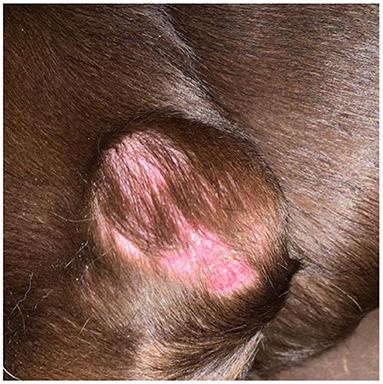
Frontiers | Case Report: Cutaneous Pleomorphic ...
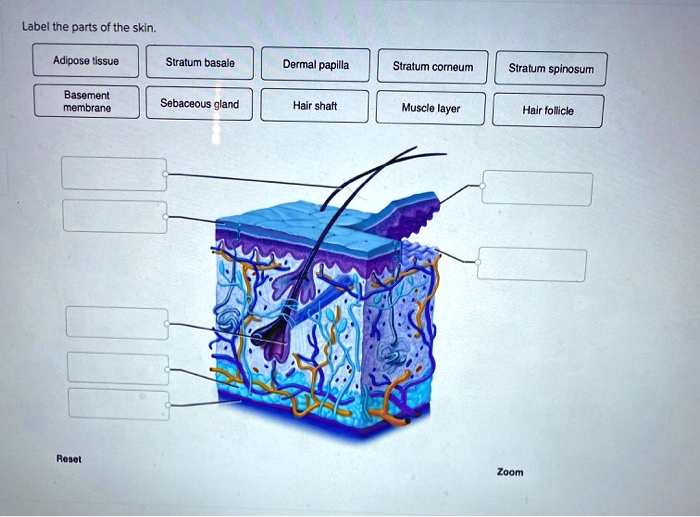
SOLVED: Label tne parts of the skin: Adipose tissue Stratum ...
4.2: Accessory Structures of the Skin - Biology LibreTexts
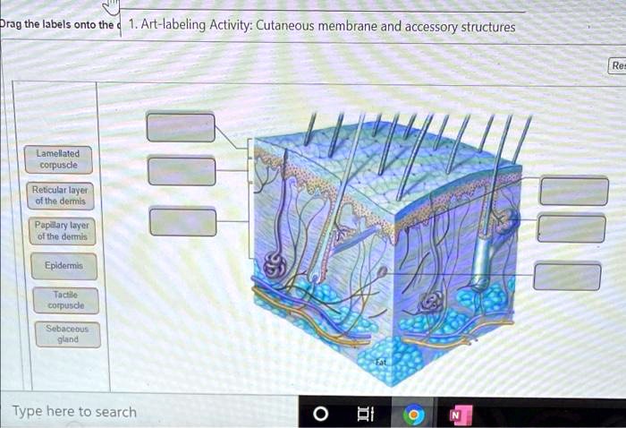
SOLVED: Drag the labels onto the 1.Art-labeling ...
Integumentary System Overview
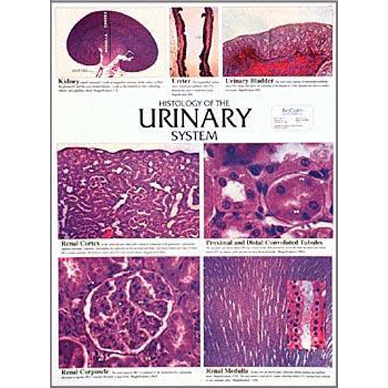
Histology of the urinary system, Chart - Southern Biological
Fine-Needle Aspiration of Solid Masses | SpringerLink
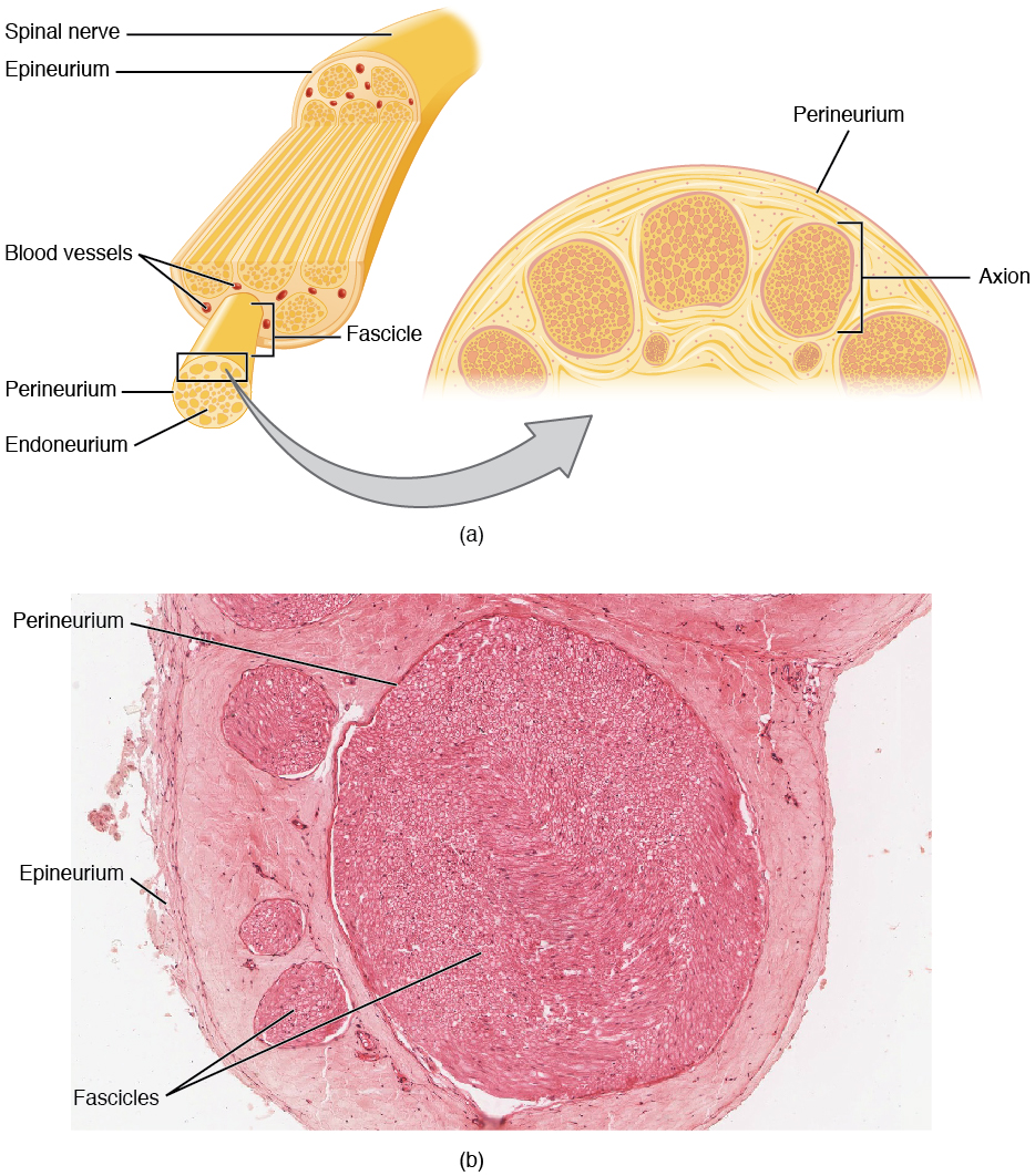
13.2 Ganglia and Nerves – Anatomy & Physiology
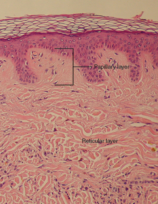
Skin and the Integumentary System

Androgen receptor messenger RNA and protein in adult rat ...

PreLab03a Integument & Prelab03b Integument Histology ...
Solved] Lab 3 Exercise 3-5 LAB 3 EXERCISE 3-5 1. In each of ...
Layers of the Skin | Anatomy and Physiology I
Chemistry Class Fabric Chemistry by Sandityche Chemistry - Etsy

Figure 7.1: Photomicrograph of Skin Diagram | Quizlet

P1: Identify Skin and Accessory Structures Diagram | Quizlet
Skin histology hi-res stock photography and images - Alamy

Blue Histology - Integumentary System
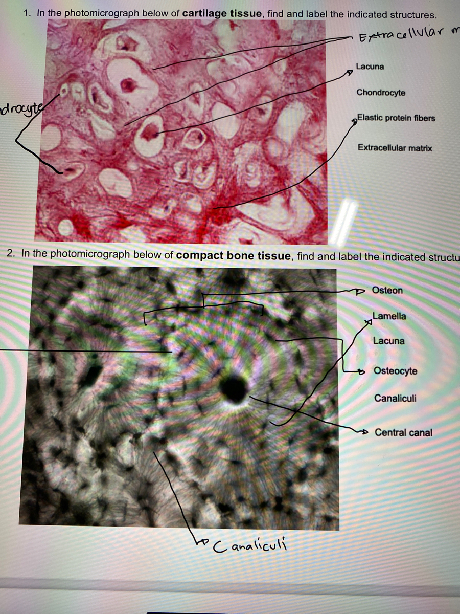
Answered: 1. In the photomicrograph below of… | bartleby

A&P 1 Exercise 7 Activity 1 & 2 & RYK and UYK.docx - LAB ...

Chapter 14 - Layers of the Skin - BIO 140 - Human Biology I ...

General Neurology | Media | MedLink Neurology
Chapter 5
Chapter 5
Hair shaft Dermal papillae Epidermis Subpapillary vascular ...
Minerals | Free Full-Text | Breaking Preconceptions: Thin ...

Diagnostic Pathology: Nonneoplastic Dermatopathology by Brian ...

Label the photomicrograph of the skin and its accessory ...

SOLUTION: Infectious diseases skin p4 - Studypool

Subungual Exostosis of the Great Toe: A Case Report and Tumor ...
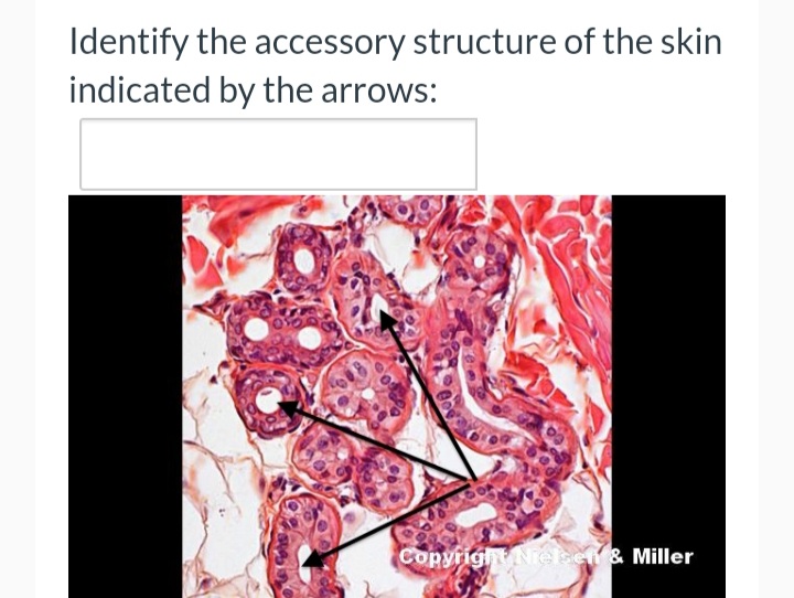
Answered: Identify the accessory structure of the… | bartleby
Integumentary System

Al mefty meningiomas by Neurocirurgiao bh - Dr Eric Grossi ...

Structural Basis of Cerebellar Microcircuits in the Rat ...

Layers of the Skin | Anatomy and Physiology I
Solved] Please help me. LAB 4 EXERCISE 4-3 82 Skin ...

Hair shaft Dermal papillae Epidermis Subpapillary vascular ...

Comparative and Functional Morphology of the Primate Hand ...

Principles of Anatomy & Physiology by Gerard J Tortora: Used ...
Label a diagram of the skin - Mrs. Sanborn's Science Class
Flat epithelium hi-res stock photography and images - Alamy
Label tne photomicrograph Of the Skin and Its accessory structures, Sebaceous gland, Duct ofl, sebaceous gland, Epidermis, Hair follicle
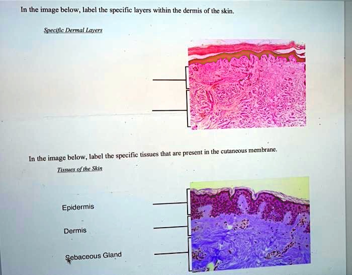
SOLVED: In the image below, label the specific layers within ...

Current considerations in medical device pathology ...
Post a Comment for "44 label the photomicrograph of the skin and its accessory structures"